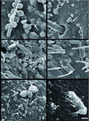
Figure 2.
Scanning electron micrographs of adult females of Peregrinus maidis infected with Beauveria bassiana CEP189 and Metarhizium anisopliae CEP 160: (a–c) B. bassiana germ tubes (arrows) penetrating through the cuticle, Bar: 1.5 µm; 2.8 µm and 2 µm, respectively, (d) M. anisopliae germ tube (arrow) entering through the hair sockets situated on forewing venation, Bar: 3.3 µm. (e) Bacillus-like bacteria (arrows) associated with two globose B. bassiana conidia on cuticle surface, Bar: 1.7 µm. (f) Bacillus-like bacteria (arrows) associated with M. anisopliae conidium on cuticle surface, Bar: 1 µm. High quality figures are available online.

