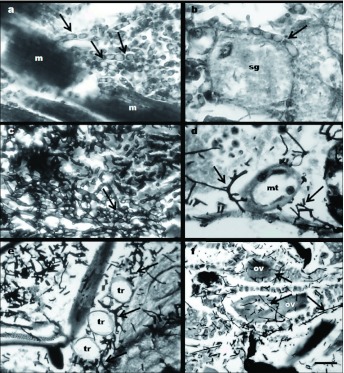
Figure 3.
Sagittal sections of Peregrinus maidis infected with Beauveria bassiana CEP189 and Metarhizium anisopliae CEP 160. (a) Hyphal bodies of M. anisopliae (arrows) in the muscle tissue (m), Bar: 8 µm. (b) Hyphal bodies of M. anisopliae (arrow) in the salivary glands (sg), Bar: 4.2 µm. (c) Hyphal bodies of B. bassiana (arrow) inside the thorax, Bar: 7 µm. (d) B. bassiana hyphae (arrows) near the Malpighian tubules (mt), Bar: 7.5 µm. (e) B. bassiana hyphae (arrows) near the tracheae (tr), Bar: 7.5 µm. (f) B. bassiana hyphae (arrows) penetrating through the ovariole wall (ov), Bar: 15 µm. High quality figures are available online.

