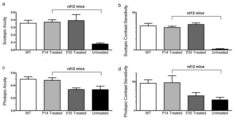
Figure 3. scAAV-RPE65 treatment restores photopic vision in rd12 mice treated at P14, and scotopic vision in rd12 mice treated at P14 or P35.
Under scotopic conditions, treatment with AAV-RPE65 at P14 (light grey bar, n=4 eyes, and at P35 (dark grey bar, n=3 eyes) leads to acuities (a) and contrast sensitivities (b) comparable to uninjected C57BL/6 mice (white bars, n=4 eyes). Untreated rd12 eyes (black bars, n=3) perform significantly poorer in tests of both scotopic acuity (a) and scotopic contrast sensitivity (b) than those from C57 or treated rd12 eyes. The level of vision in these untreated eyes is equivalent to no visual function. Under photopic conditions, (c) rd12 mouse eyes treated with AAV-RPE65 at P14 (light grey bar, n=4) have significantly better acuity than untreated rd12 control eyes (black bar, n=3) and maintain photopic acuity comparable to wild type C57BL/6J eyes (white bar, n=4), while eyes treated at P35 (dark grey bar, n=3 eyes) show no significant change from their untreated rd12 counterparts (black bar, n=3 eyes). (d) As with photopic contrast sensitivity, rd12 eyes treated at P14 (light grey bar, n=4 eyes), but not those treated at P35 (dark grey bar, n=3 eyes) exhibit photopic contrast sensitivity near wild type C57BL/6J levels (white bar, n=4 eyes).

