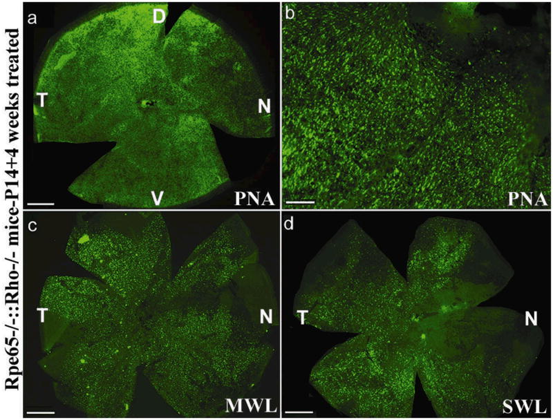
Figure 7. Retinal whole mounts of Rpe65−/−::Rho−/− mice 4 weeks after vector treatment at P14 and stained with PNA, MWL-cone opsin antibody or SWL-cone opsin antibody.
PNA staining revealed that cones were abundant in the peripheral (a) and central (b) regions of a P14 treated Rpe65−/−::Rho−/− retina. These cones were positive for both MWL cone opsin (c) and SWL cone opsin (d). D: dorsal; V: ventral; T: temporal; N: nasal. Scale bars (a, c, d) = 500 μM. Scale bar (b) = 100 μM.

