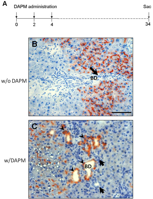
Figure 2.

Appearance of DPPIV in bile ducts cells after repeated DAPM administration (DAPM × 3).(A) Schematic representation of repeated DAPM administration protocol. DAPM (50 mg/kg) administered at day 0, 2, and 4 to the DPPIV chimeric rats. Rats sacrificed at day 30 after the last DAPM injection. DPPIV staining before (B) and after (C) repeated DAPM administration to the DPPIV chimeric rats. Arrowheads point to the DPPIV positive bile ducts. Arrows indicate DPPIV negative bile ducts. The number of DPPIV positive bile ducts was determined after counting DPPIV positive bile ductules in liver sections obtained from different lobes of liver from 3 individual rats separately. None of the bile duct cells of the DPPIV chimeric rats were positive before DAPM treatment. ~20% bile ducts were noted to be DPPIV positive after DAPM × 3 protocol. Scale bar = 100 μm.
