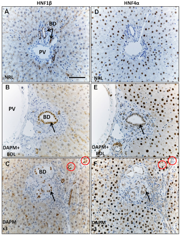
Figure 5.
HNF1β and HNF4α immunohistochemistry on serial liver sections. (A) normal control rats (NRL, normal rat liver), (B) rats that underwent DAPM + BDL treatment, or (C) repeated DAPM treatment (DAPM × 3). HNF1β and HNF4α coexpressing cells are pointed by an arrow. HNF1β positive but HNF4α negative bile ducts pointed by circles. PV, portal vein; BD, bile duct. Scale bar = 100 μm.

