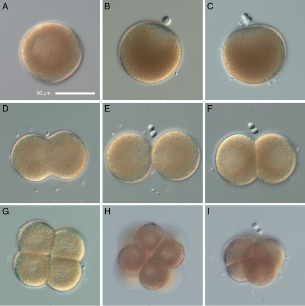
Figure 1.
Early cleavage in Micrura alaskensis. DIC images. A. Unfertilized egg undergoing germinal vesicle breakdown. B. Fertilized egg with the first polar body. C. Formation of the second polar body. D-F. First cleavage. D. Broad initial cleavage furrow. E. There is no egg chorion, and the blastomeres separate almost completely. F. Embryo re-compacts after first cleavage. G. Polar view of the four-cell stage. H. Vegetal view of an eight-cell stage illustrating dextral spiral cleavage. I. Side view of an eight-cell stage. Apical pole, marked by polar bodies, is up. Animal micromeres are larger than the vegetal macromeres.

