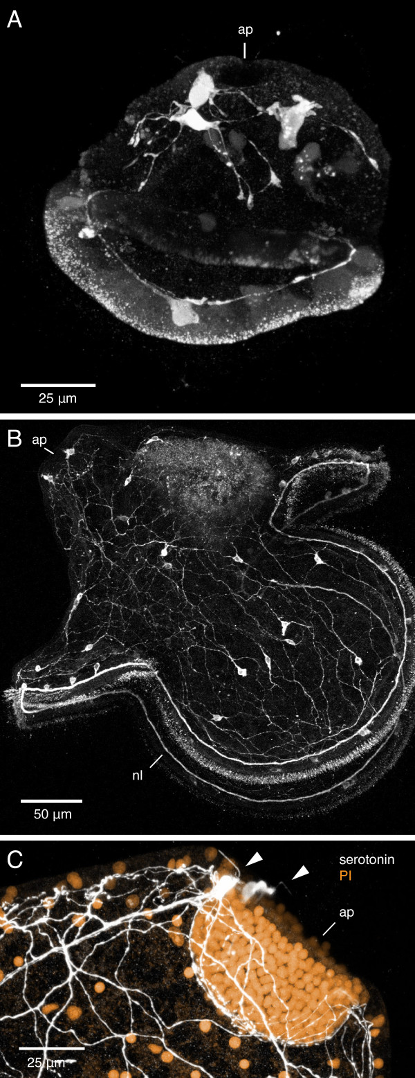
Figure 10.
Serotonergic nervous system of the pilidium larva of Micrura alaskensis. Confocal projections. A. Z-projection of a 40-hour-old pilidium larva stained with anti-5HT antibody. Apical plate (ap) is up. Serotonergic neurons are present both in the apical region and along the ciliated band. B. Z-projection of a month-old pilidium larva (head and trunk stage) stained with anti-5HT antibody. Note the extensive serotonergic network, a pair of neurons associated with the apical plate, and the marginal ciliary nerve (nl) associated with the larval ciliated band. C. Z-projection of the apical plate of a month-old pilidium larva stained with anti-5HT antibody (white) and propidium iodide (orange). Note the two serotonergic neurons (arrowheads) associated with the apical plate.

