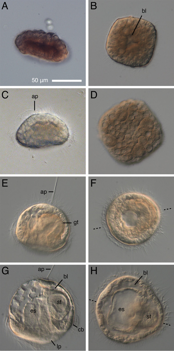
Figure 2.
Development of Micrura alaskensis from blastosquare to young pilidium. DIC images. Left panels - side view, right panels - polar view. A-B, D. Ciliated blastosquare (18-20 hours after fertilization). Note the small blastocoel (bl) on B and spiral cleavage pattern apparent in a slightly compressed blastosquare (D). C. Gastrula with an apical tuft (ap) (27-hours-old). E-F. Two-day-old pilidium (49-53-hours-old) showing bilaterally symmetrical gut (gt) and a long apical tuft. Plane of bilateral symmetry is indicated by dashed line on F and H. G-H. Three-day-old pilidium has a blind gut differentiated into esophagus (es) and stomach (st), a prominent apical tuft, ciliated band (cb), and nascent lateral lappets (lp). The young pilidium increases in size even before onset of feeding due to the expansion of the blastocoel.

