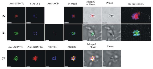
Figure 9.
Late schizonts show a central concentration of PfSHMTc fluorescence. (A) Post-mitotic schizont showing a concentration of PfSHMTc fluorescence in the centre of the parasite, and overlapping the outer zone of haemozoin. PfSHMTc is largely excluded from the nuclei. (B) Post-mitotic schizont showing a concentration of PfSHMTc fluorescence in the centre of the parasite as well as at low intensity in the multiple small apicoplasts. Note the merozoite buds arranged in a radial pattern centred on the future residual body. (C) A post-mitotic parasite probed with both anti-PfSHMTc (IgY) and anti-PfSHMTm (IgG). Both SHMT proteins show a similar, but not identical distribution, as described for image series (A) and (B) above (scale bars 3 μm).

