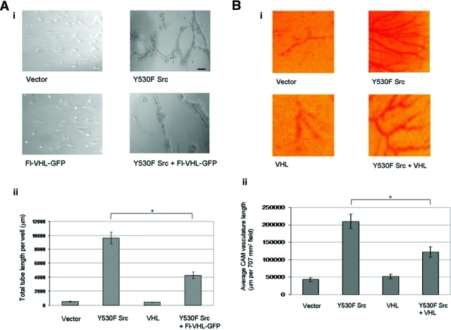
Figure 6.

Src destabilization of von Hippel–Lindau (VHL) protein upregulates tubulogenesis in vitro and chick embryo chorioallantoic membrane blood vessel growth. (A) (i) Photographs of endothelial tube formation assays, on conditioned media collected from HEK 293T cells transfected with the indicated plasmids 48 h posttransfection. (ii) Quantification of capillary network formation was done by measuring total tube length per well using ImageJ image data analysis software. Scale bar: 100 µm. The graph charts the averages ± 1 SD from 3 separate transfections done in duplicate. Statistical significance was determined by Student t test; *P < 0.05. (B) (i) Photographs of developing chick chorioallantoic membranes (CAMs), implanted with gelatin sponges loaded with HEK 293T cells transfected with the indicated plasmids. (ii) Quantitation of angiogenesis was done by measuring the length of vasculature per 707 mm2 field, using ImageJ image data analysis software. Four fields per CAM were measured for 9 CAMs per transfection condition. The graph charts the averages ± 1 SD. Statistical significance was determined by Student t test; *P < 0.05.
