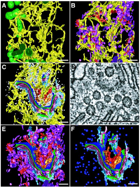
Figure 3.
Structural evidence for physical relationships among different organelles in the modeled region. The ER is closely apposed to mitochondria (dark green) (A), clathrin-positive (red) and clathrin-negative (purple) endo-lysosomal compartments (B). (C) Here, 2,119 small (average diameter 52 nm), spherical, non-clathrin vesicles (white) were distributed close to the Golgi and ER. (D) Higher magnification image extracted from tomographic data showing the numerous tethers connecting small vesicles to each other and to Golgi membranes. Bar = 250 nm. (E) Subsets of endo-lysosomal compartments with distinct morphological profiles were clustered together in the Golgi region. (F) Here, 132 dense core vesicles (bright blue; average diameter 100 nm) were present in the Golgi region but were apart from the Golgi stack. Bars = 500 nm.

