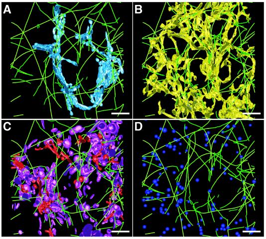
Figure 4.
The MT cytoskeleton associates with membranes of the Golgi, ER, and endo-lysosomal compartments. The paths of MTs (bright green) closely followed and occasionally formed contacts with the membranes of C1 (light blue; A) and the ER (yellow; B). Clathrin-positive (red) and clathrin-negative (purple) compartments (C) and dense core vesicles (bright blue; D) in the reconstructed volume were modeled so that in situ spatial relationships between these elements and MTs could be reliably quantified and assessed. Bars = 500 nm.

