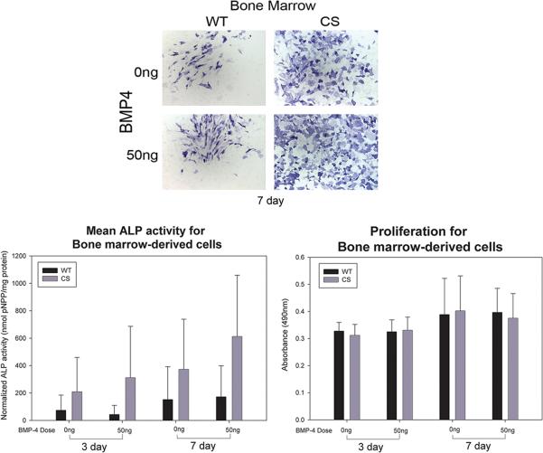
Figure 3.
A) Histophotomicrographs showing ALP-stained (blue) bone marrow-derived cells isolated from wild-type (WT) or craniosynostotic (CS) rabbits after 7 days in culture. Bone marrow-derived cells derived from CS rabbits showed more ALP staining than WT counterparts. B) Graph depicting the mean quantified ALP activity (error bar depicts standard deviation) of bone marrow-derived cells from WT (n=9) or CS (n=18) rabbits. Analysis showed significant differences between WT and CS cells (p<0.001) and between 3 and 7 days (p<0.05). C) Graph depicting the means and standard deviations of absorbance collected as a measure of total cell number. No statistical differences were identified due to BMP4 or donor diagnosis after either 3 or 7 days in culture.

