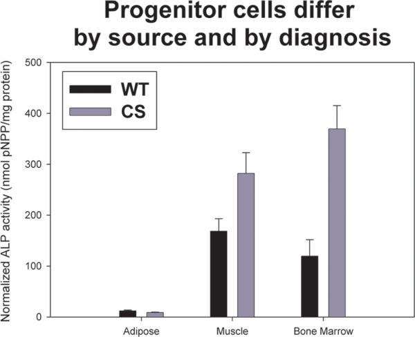
Figure 4.

Graph showing the mean and standard errors for quantitative ALP activity for adipose-, muscle-, and bone marrow-derived cells from either wild-type (WT) or craniosynostotic (CS) rabbits (error bar depict standard error). Statistical analysis showed a significant cell type by phenotype interaction (p<0.001), suggesting that cells derived from WT and CS rabbits were different from each other. For WT rabbits, cells derived from muscle showed the most ALP activity, whereas in CS rabbits, cells derived from bone marrow had the highest ALP activity. Adipose-derived cells were similar between WT and CS rabbits.
