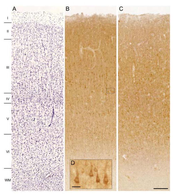
Figure 5.
Brightfield photomicrographs showing Nissl staining (panel A) and laminar distribution pattern of OXSR1 immunoreactivity in human DLPFC area 9 for a subject with a short PMI (5.5 hours, panel B) and a long PMI (19.3 hours, panel C). The insert shows a magnification of OXSR1 immunoreactivity in layer 3.

