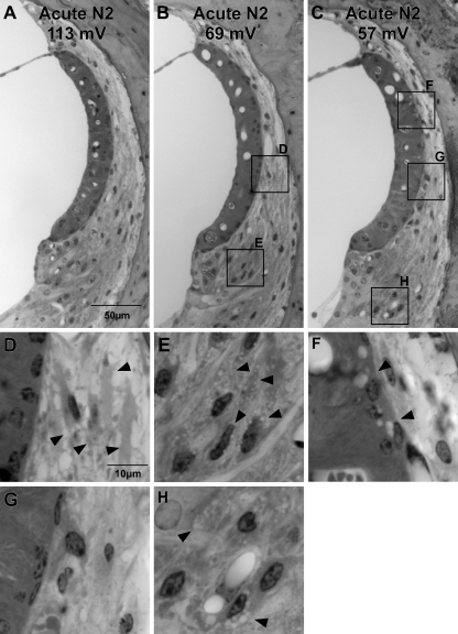
FIG. 11.
Examples of the three major patterns of cellular appearance observed in the cochlear upper basal turn 1–3 h after noise in B6/BALB N2 backcross mice. EPs are shown for each animal. A Normal EP attended by minimal pathology, as detected by light microscope. B Mouse with reduced EP combining minimal strial pathology with clear pathology of type I fibrocytes (expanded in (D), arrowheads) and type II fibrocytes (expanded in (E), arrowheads). C Mouse combining clear strial pathology (expanded in (F), arrowheads) with pathology of type II fibrocytes (expanded in (H), arrowheads), but showing minimal pathology of type I fibrocytes (expanded in (G)).

