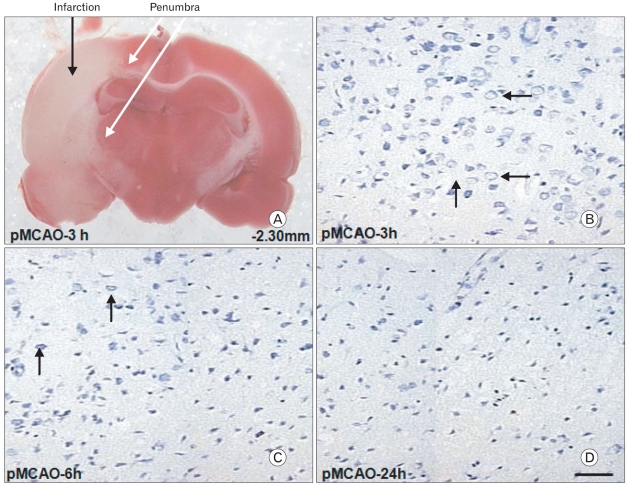
Fig. 1.
TTC (2,3,5-triphenyltetrazolium chloride) staining of brain slice from bregma -2.30 mm 3 h after permanent middle cerebral artery occlusion (pMCAO). Tissues of the penumbra (marked with white arrows) represented the red zone near the infarction zone in the ipsilateral hemisphere over a series of brain sections (A). Cresyl violet staining demonstrated that viable cells in the penumbra (marked with short black arrows) were significantly decreased 3 to 6 h after pMCAO (B, C). Moreover, viable cells were not detected 24 h after pMCAO which resembled the ischemic core (D). Scale bar=50 µm.

