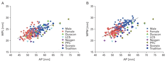
Fig. 5.
Comparison of the width of the resected posterior femoral medial condyle (A) and posterior femoral lateral condyle (B) with that of femoral designs for matching AP dimensions. The line represents the average values for the female and male population. All designs show undercoverage for both condyles.

