
Fig. 5.
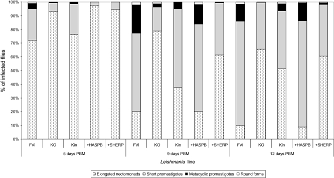
Morphological forms of the lines described in Fig. 3 during development in P. papatasi. The guts of L. major infected females were sampled at 5, 9 and 12 days PBM and parasite morphometry determined as described (Experimental procedures). The % of each form found in infected flies at each time point is shown. Differences among lines were significant and increased from day 5 PBM (P < 0.0001, χ2 = 93.266, d.f. = 12) to day 9 PBM (P < 0.0001, χ2 = 246.947, d.f. = 12) and day 12 PBM (P < 0.0001, χ2 = 283.947, d.f. = 12).