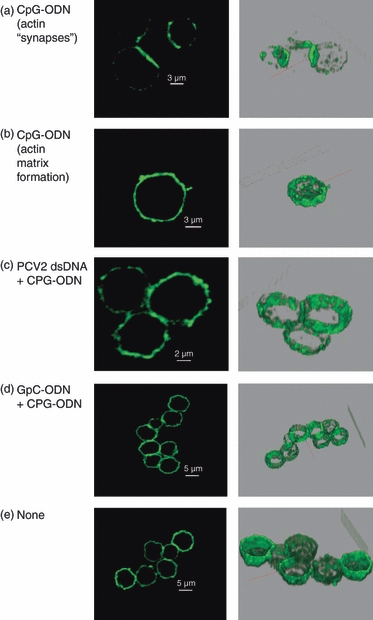
Figure 3.

Confocal microscopy. Sectional scanning of plasmacytoid dendritic cells showing actin staining. Cells were isolated by cell fractionation on a 55% Percoll gradient and CD4+ selection. Cells were incubated fro 30 min with 100 ng porcine circovirus type 2 (PCV2) DNA, 10 μg/ml GpC, or untreated (none) before stimulation with 10 μg/ml CpG. Cells were then fixed and permeabilized and stained for actin with phalloidin. (a) CpG-oligodeoxynucleotide (ODN) stimulation of microfilament re-organization seen in terms of synaptic-like actin formations between cells. (b) CpG-ODN stimulation cytoplasmic microfilament matrix formation. (c, d) PCV2 double-stranded DNA and GpC treatment prevents CpG-ODN-induced microfilament re-organization (no ‘synaptic’ or cytoplasmic matrix formation). (e) Untreated. The right column shows three-dimensional images of cells sections. The data are representative of two independent experiments.
