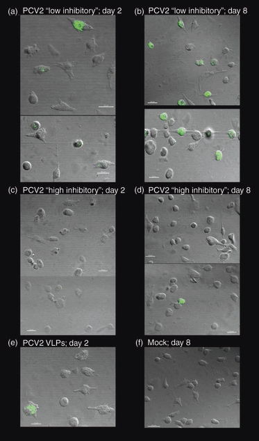
Figure 6.

Monocyte-derived dendritic cell endocytosis: confocal microscopy. Monocyte-derived dendritic cell were incubated with porcine circovirus type 2 (PCV2) low (a, b) or high (c, d) inhibitory stocks at a multiplicity of infection of 0.1 of the 50% tissue culture infective dose/cell and incubated for 2 days (a, c) or 8 days (b, d) at 39°. Cells were washed and stained for the PCV2 capsid antigen. A positive control (100 ng of PCV2 virus-like particle; VLP) stained after 2 days is shown in (e), and a negative control (mock cell lysate) stained after 8 days of incubation is shown in (f). The data are representative of two independent experiments.
