
Figure 3.
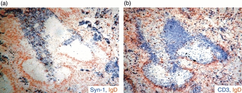
Extrafollicular (EF) foci in Trypanosoma cruzi-infected mice infiltrate T-cell zone Photomicrograph of serial sections from the same spleen obtained from 11-day T. cruzi-infected mice stained with: anti-CD138, anti-IgD and anti-CD3. (a) CD138+ EF focus stained blue and IgD+ B cells (follicular mantle) stained brown; (b) CD3+ T cells in blue and the follicular mantle in brown.