
Figure 2.
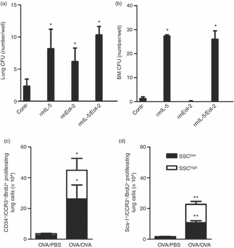
Haematopoietic activity of interleukin-5 (IL-5) and eotaxin-2 and newly produced eosinophil-lineage-committed cells proliferate after allergen exposure. (a) Colony-forming units (CFUs) after 8–14 days of culture of lung CD34+ cells and (b) 8 days of culture of bone marrow (BM) CD34+ cells harvested from ovalbumin (OVA) -sensitized/exposed mice (n = 3–6) *P< 0·05 for comparison between control and cytokine/chemokine-stimulated colonies; Wilcoxon signed rank test. The number of CCR3+ BrdU+ proliferating cells in the lung, analysed by FACS of magnetically enriched CD34+ cells (c) and Sca-1+ cells (d) from OVA-sensitized/exposed BALB/c mice compared with OVA-sensitized but PBS-exposed mice. Open bars represent cells gated on the SSChigh cell population and dark bars are cells gated on the SSClow cell population. Data are shown as mean (+ SEM) (n = 5–7). *P< 0·05 and **P< 0·01 between OVA-exposed and PBS-exposed groups.