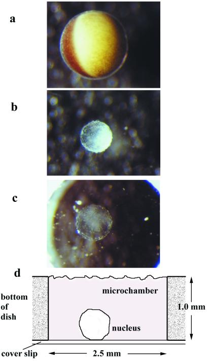
Figure 1.
Preparation of an isolated Xenopus oocyte nucleus for transport measurement. The nucleus of a stage VI Xenopus oocyte (a) was isolated manually (b) and, after manual purification with a fine-glass needle, deposited in a microchamber (c). The microchamber (d) had a volume of 5 μl. Transport was monitored by confocal scans by using either an inverted or an upright setup.

