Figure 1.
Expression Pattern of KASI.
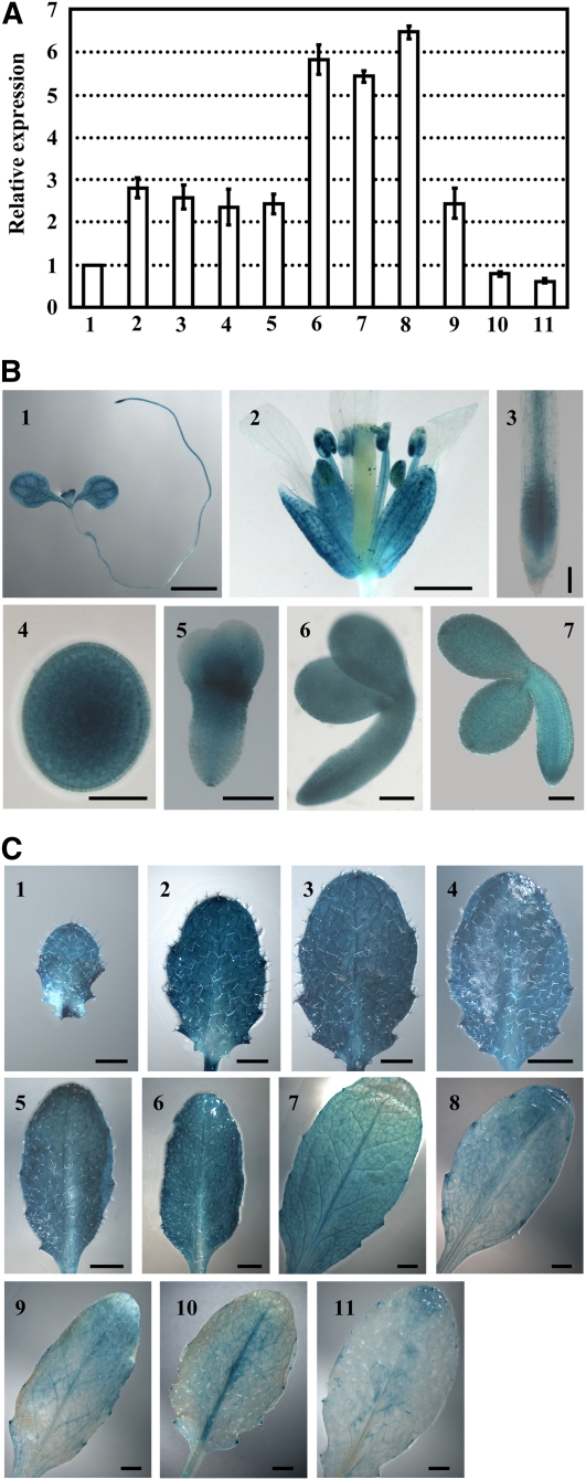
(A) qRT-PCR analysis of KASI expression in various tissues. The relative expression of the KASI gene was detected in 7-d-old seedling (2) and root (3), flower (4), young silique (<0.5 cm, 5), embryos at late cotyledon stage (6), the 8th or 7th rosette leaf at 16 DAG (7 and 8), and the 7th rosette leaf at 23, 28, or 35 DAG (9 to 11) by setting the expression of KASI in stalk (1) as 1.0. The data were from three biological replicates and presented as means ± sd.
(B) Promoter-reporter gene (GUS) fusion studies reveal the expression of KASI in 7-d-old seedling (1), flower (2), root (3), and embryos at different developmental stages (4 to 7). KASI expression in embryos at the globular stage (4), heart-shaped stage (5), early cotyledon stage (6), and later cotyledon stage (7) are analyzed. Bars = 2 mm in (1), 1 mm in (2), 10 μm in (4), and 100 μm in (3) and (5) to (7).
(C) Promoter-GUS fusion studies reveal the KASI expression in rosette leaves at different developmental stages. Seventh rosette leaves at 15 (1), 17 (2), 19 (3), 21 (4), 23 (5), 25 (6), 27 (7), 29 (8), 31 (9), 33 (10), and 35 DAG (11) were used for analysis. Bars = 1 mm in (1) to (3) and 2 mm in (4) to (11).

