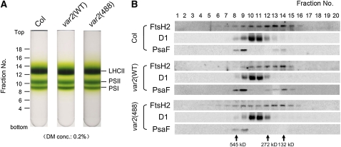
Figure 4.
Sucrose Density Gradient Analysis of Thylakoid Membrane Proteins.
(A) Photographs of 0.1 to 1.3 M sucrose density gradients. Thylakoid membranes from Col, var2(WT), and var2(488) were solubilized using 0.2% DM. Relative positions of the fractions and the locations of LHCII monomer/trimer (LHCII), PSII monomer (PSII), and photosystem I complex (PSI) are indicated.
(B) Immunoblot analysis of the sucrose density gradient fractions. The fractions were collected from the tubes of the sucrose density gradients shown in (A). Equal amounts of the fractions were subjected to SDS-PAGE and probed with anti-FtsH2, anti-D1, and anti-PsaF. Immunoblot analysis data from Col, var2(WT), and var2(488) are shown.

