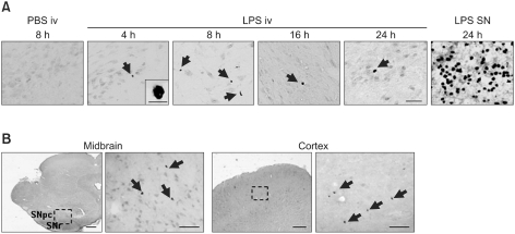
Figure 2.
Neutrophils infiltrate the brain in response to iv LPS administration. (A) Sections were obtained from the midbrain at the indicated times after LPS injection (LPS iv), or 24 h after direct LPS injection into the SN (LPS SNpc), and stained with an anti-MPO antibody. PBS-injected brain sections were used as positive controls. (B) Brain sections were obtained from the midbrain and the cortex 8 h after iv LPS injection, stained with an anti-MPO antibody, and MPOexpression was visualized using a peroxidase-conjugated secondary antibody. Scale bars: 200 µm (left panels in B); 50 µm (A and right panel of each region in B); and 10 µm (inset in A).

