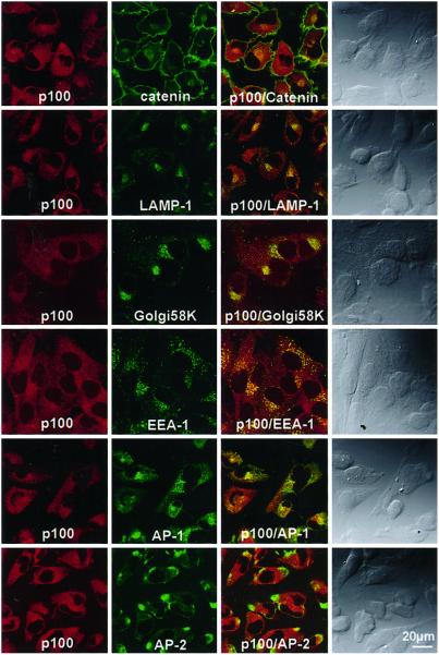
Figure 8.
Confocal images of T98G cells stained for ARF-GEP100 and organelle marker proteins. Cells were reacted with rabbit anti-ARF-GEP100 (p100) antibody and mouse monoclonal antibody against β-catenin, LAMP-1, Golgi 58K protein, EEA-1, AP-1, or AP-2, followed by Texas Red-labeled anti-rabbit IgG and FITC-labeled anti-mouse IgG. In the first two columns are pairs of images of the same cells. Images in the third column are superimposed images of the preceding panels. In the fourth column are Nomarski images.

