Fig. 1.
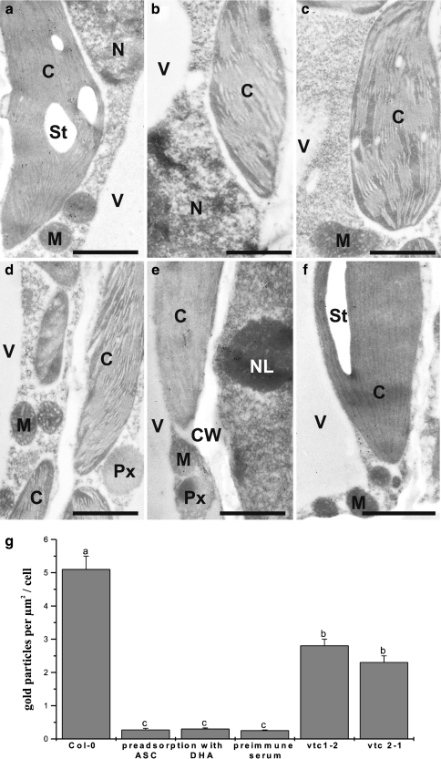
Specificity of immunocytochemical ascorbate labeling. Transmission electron micrographs (a–f) and quantitative analysis (g) showing the overall distribution of gold particles bound to ascorbate in leaf cells of Arabidopsis Col-0 plants (a–d) and the Arabidopsis mutants vtc1-2 (e) and vtc2-1 (f). a Ascorbate-specific labeling detected in different cell compartments of Arabidopsis Col-0 plants such as chloroplasts (C), mitochondria (M), nuclei (N), and vacuoles (V). Negative controls include preadsorption of the anti-ascorbate antisera with ascorbic acid (ASC, b), dihydroascorbate (DHA, c), and the treatment of sections with preimmune serum instead of the primary antibody (d). Very few or no gold particles were present on these sections. Gold particle density was significantly lower in cells of the vtc1-2 (e) and vtc2-1 (f) mutants. CW cell walls, NL nucleoli, Px peroxisomes, St starch. Bars 1 μm. g Significant differences between the samples are indicated by different lowercase letters; samples which are significantly different from each other have no letter in common. P < 0.05 was regarded significant analyzed by the Kruskal–Wallis test, followed by post hoc comparison according to Conover

