Figure 3.
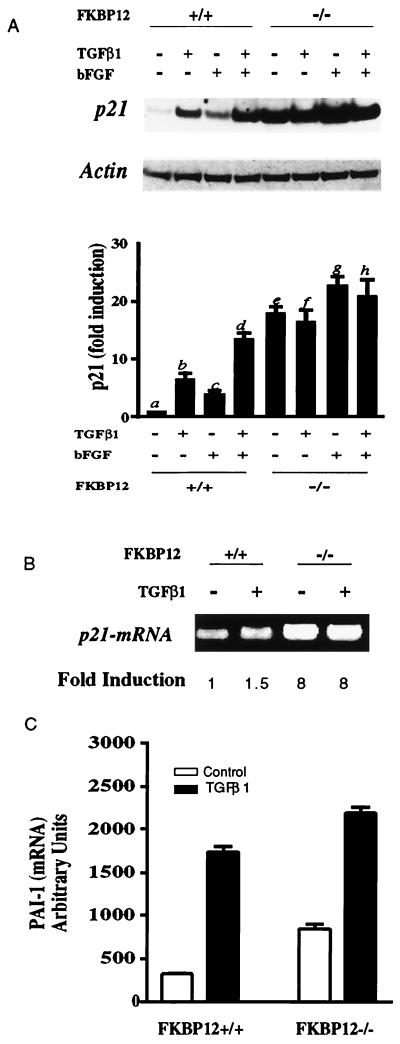
Increased p21(WAF1/CIP1) expression in FKBP12−/− cells. (A) TGF-β1 and bFGF treatments for 24 h induce p21 protein in FKBP12+/+ cells (bars b–d are different from bar a, P < 0.01) and their effects are additive (bar d is different from bars b and c, P < 0.01). In FKBP12−/− cells, p21 protein is greatly augmented (bar e is different from bar a, P < 0.001), and TGF-β1 does not elicit any further increase although bFGF does augment p21 (bar g is different from bar e, P < 0.05). Data are the mean ± SEM of five experiments. (B) p21 mRNA is augmented in FKBP12−/− fibroblasts. p21 mRNA was monitored by real-time PCR and RT-PCR; RNA was extracted from FKBP12+/+ and FKBP12−/− cells. p21 mRNA was 8-fold higher in FKBP12−/− cells. TGF-β1 treatment (3 h) induced p21 in FKBP12+/+ cells but not in FKBP12−/− cells. Data are representative of data from three experiments, whose results varied less than 15%. (C) PAI-1 mRNA is augmented in FKBP12−/− fibroblasts. PAI-1 mRNA was monitored by real-time PCR and RT-PCR; RNA was extracted from FKBP12+/+ and FKBP12−/− cells. PAI-1 mRNA was 2.5-fold higher in FKBP12−/− cells than in FKBP12+/+ cells. However, consistent with previous reports (12, 23), TGF-β1 treatment (3 h) induced PAI-1 in both FKBP12+/+ cells and FKBP12−/− cells by 5- and 3-fold, respectively. Data are representative of data from three experiments, whose results varied less than 15%.

