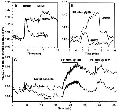
Figure 6.
Imaging of cGMP in Purkinje neurons. Emission ratios (483/535 nm) indicating cGMP concentrations were imaged in cygnet-2-transfected cerebellar Purkinje neurons in organotypic culture. (A) cGMP responses to 0.1 mM NONO (indicated by the bar) without and with 0.6 mM IBMX (−IBMX and +IBMX). Both traces are from the soma of the same cell. (B) cGMP transients responses to 4 Hz parallel fiber stimulation (duration indicated by the bar) without and with IBMX in the medium. Both traces are from the same cell. (C) Comparison of cGMP responses to different frequencies and duration of parallel fiber stimulation in the soma and distal dendrites.

