Figure 2.

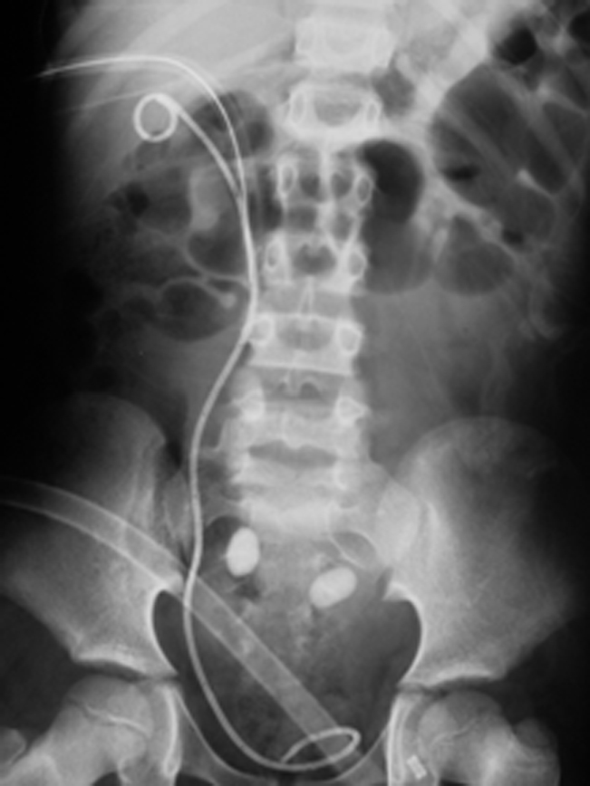
X-ray KUB showing 2 calculi overlying the sacrum in the left ectopic pelvic kidney. Nephrostomy tube and double “J” in situ seen on the right side.

X-ray KUB showing 2 calculi overlying the sacrum in the left ectopic pelvic kidney. Nephrostomy tube and double “J” in situ seen on the right side.