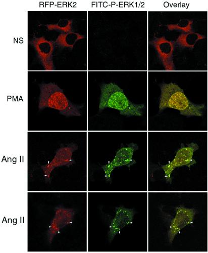
Figure 2.
Effect of angiotensin II on the cellular distribution of RFP-ERK2 and phospho-ERK1/2. HEK-293 cells were transiently transfected with plasmid DNA encoding HA-AT1aR, Flag-β-arrestin-2, and RFP-ERK2. Serum-starved cells were treated with vehicle (NS), PMA, or angiotensin II (Ang II) for 15 min, fixed with paraformaldehyde, permeabilized, and stained with FITC-conjugated monoclonal anti-phospho-ERK1/2 before examination by confocal microscopy. The distribution RFP-ERK2 (red) and FITC-stained phospho-ERK1/2 (green) are shown in the single channel images. Colocalization of RFP-ERK2 and phospho-ERK1/2 is shown in the overlay images (yellow; arrows). Each image depicts a representative confocal microscopic image from one of three separate experiments.

