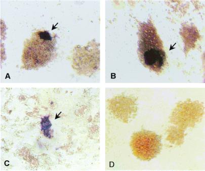
Figure 3.
Distribution of vasa-positive embryo cells. Cultures maintained for 3 days (a) and 8 days (b) in RTS34st cell-conditioned medium or 3 days on RTS34st feeder cells (c) were examined by in situ hybridization by using a vasa-specific antisense probe. The control culture (d) was grown for 8 days in conditioned medium and hybridized with sense probe. [Magnification = ×200 (a, b, and d), and = ×100 (c).]

