Figure 3.

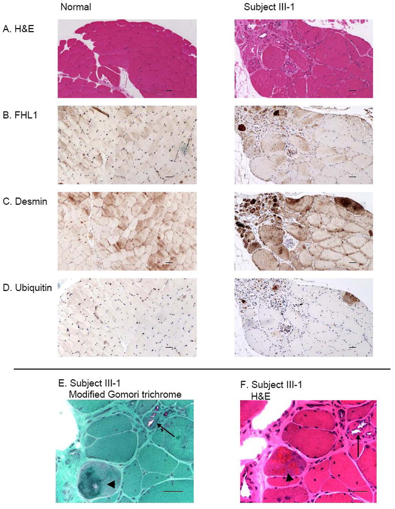
Muscle biopsy from a subject III-1 in the family with FHL1 mutation revealing myopathy and FHL1 inclusions that are desmin and ubiquitin-positive. In muscle from the proband, H&E staining (A and F) reveals that muscle fiber size is markedly variable with numerous small rounded atrophic fibers and enlarged hypertrophic fibers. Central nuclei are increased in number and some fibers contain multiple central nuclei. Scattered intrasarcoplasmic eosinophilic inclusions are present in some fibers. In muscle from a normal control, muscle fiber size is generally equal and nuclei are peripherally placed and central nuclei are uncommon. There is no increase in fibrous connective tissue. Immunohistochemistry for FHL1 (B), desmin (C) and ubiquitin (D) shows abnormal dense staining present in some atrophic fibers. In a normal control, normal desmin-positive striations are present; no ubiquitin or FHL1 staining is observed. Rimmed vacuoles (arrow) and eosinophilic inclusions (arrowhead) are revealed more clearly at higher magnification in E (modified Gomori trichrome stain) and F (H&E). Scale bars: 100 μm.
