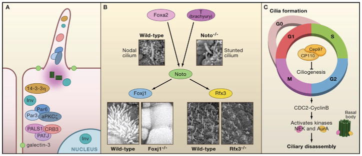
Figure 3. Control of Ciliogenesis.
(A) Ciliary proteins localize to cell junctions and polarity proteins localize to the primary cilium. Inversin localizes to three separate cellular protein pools: the primary cilium, cell junctions, and the nucleus. Two different polarity complexes, Par3/Par6/aPKCζ and CRB3/PATJ1/PALS, as well as the polarity protein 14-3-3η, localize to cell junctions and the primary cilium. Galectin-3 also marks a membrane region of increased lipid density surrounding the cilium.
(B) The transcription factors Noto, FoxJ1, and Rfx3 play a role in ciliogenesis. Noto acts downstream of Foxa2 and T (brachyury) and upstream of FoxJ1 and Rfx3. Embryonic node and nodal cilium in E7.5 wild-type and Noto-deficient mice (Beckers et al., 2007) reveal structurally abnormal and stunted cilia. Images are reproduced with permission from PNAS, Vol. 104, p. 15765–15770. Copyright (2007) National Academy of Sciences, U.S.A. Scanning electron micrographs of explanted trachea show multiciliated cells covering most of the apical surface in wild-type trachea. These are absent from mice that are Foxj1 deficient, whereas primary cilia are unaffected. Scanning micrographs of embryonic nodes of Theiler stage 11c of wild-type and Rfx3 deficient mice. Cilia in Rfx3 deficient mice are shorter and misoriented compared to the wild-type (Bonnafe et al., 2004). Images reproduced from Molecular and Cellular Biology, 2004, Vol. 24, p. 4417–4427, with permission from the American Society for Microbiology.
(C) A complex of Cep97 and CP110 suppresses ciliogenesis throughout the cell cycle. Mitotic entry is regulated by the G2/M transition through CDC2-Cyclin B, which activates several kinases, including the Nek family and AurA. In murine inner medullary collecting duct (IMCD3) cells, NEK5 localizes to the centrosome and the distal tip of the basal body. In human retinal pigment epithelial cells, a pool of AurA localizes to the basal body.

