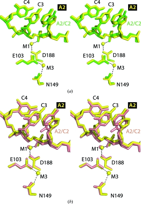
Figure 4.
Stereo images of active-site superpositions of structures with secondary-site DNA. (a) Superpositions of active site A of SgrAI bound to uncleaved primary-site DNA and Ca2+ (yellow) and bound to secondary-site DNA and Ca2+ (green) using the Cα atoms of residues 149, 188 and 103. Dashes indicate ligations to the metal ions. (b) As in (a) but with SgrAI bound to secondary-site DNA and Mg2+ (salmon).

