
Figure 11. Disrupted actin organization in lenses of Dlg-1 mutant mice.
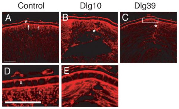
Longitudinally oriented, cryosections of eyes from control (A, D), Dlg10 (B, E) and Dlg39 (C) P2 mice were stained with phalloidin to visualize filamentous actin. Shown are representative sections. A, D: In controls, actin is concentrated at the basal and apical surfaces of epithelial cells. At the apical ends of the fiber cells actin has a uniform pattern and is highly concentrated. B, E: In lenses from Dlg10 mice, actin accumulation in the epithelium appeared normal. However, in the fibers actin organization was disrupted where anterior sutures normally form (asterisk). C: In lenses from Dlg39 mice, actin accumulation was decreased in the apical ends of the fiber cells (box) and actin organization was nonuniform, especially where the suture forms. e=epithelium, f=fibers. Scale bars=50μm.