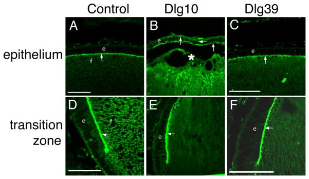
Figure 12. Mislocalization of the apical polarity marker, ZO-1, in the central epithelium of lenses of Dlg10 embryos.
Longitudinally oriented, paraffin embedded eye sections from E17.5 control (A,D), Dlg10 (B,E) and Dlg39 (C,F) embryos were immunostained with anti-ZO-1 antibodies. A: Immunostained control lenses (A), showed ZO-1 localized exclusively to the apical membrane of the cells in the central epithelium and apical tips of the fiber cells (arrow). Immunostained lenses of Dlg10 embryos (B) showed ZO-1 on all membranes of central epithelial cells and showed discontinuous staining on the apical membrane (arrows). C: Immunostained lenses of Dlg39 embryos showed staining in the epithelium that was indistinguishable from that of control lenses. D-F: In the transition zone, immunostaining showed localization of ZO-1 at the apical surface of epithelial cells in lenses from control mice (D) as well as in lenses from Dlg10 (E) and Dlg39 (F) mice (arrows). e=epithelium, f=fibers, asterisk=fiber cell elongation defect. Scale bars=50μm.

