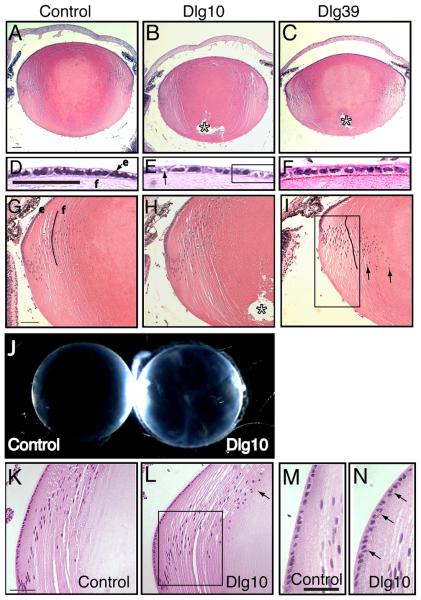
Figure 3. Morphological defects and cataract formation in lenses from postnatal Dlg-1 mutant mice.
Longitudinally oriented, paraffin embedded eye sections from control, Dlg10 and Dlg39 postnatal mice were stained with hematoxylin and eosin. Shown are representative stained sections. Whole lenses from control and Dlg10 mice were also examined for cataract formation. A-C: Low magnification view of lenses from control (A), Dlg10 (B) and Dlg39 (C) P2 mice. Posterior suture defects are readily apparent in the lenses of Dlg10 and Dlg39 mice (asterisk). D-F: Higher magnification view of the epithelium from control (D), Dlg10 (E) and Dlg39 (F) P2 mice. The epithelium from the lenses of the Dlg10 mice show irregularities in the organization and appearance of the nuclei, vacuoles (arrow) and regions of loosely packed cells lacking nuclei (box). G-I: Higher magnification view of the transition zone in lenses of the control, Dlg10 and Dlg39 P2 mice. In (I) the eosin staining was weaker, suggesting defects in the packing of the fibers in Dlg39 mice (I, Box). Also, nuclei did not bow upwards, but instead extended into the posterior region of the lens (arrows). Additionally, fibers did not maintain their normal “C” shape (lines). e=epithelium, f=fibers. J: Whole lenses from control and Dlg10 P24 mice were viewed under a dissecting microscope. Lenses from Dlg10 mice exhibited cataracts. K-L: View of the bow region from control (K) and Dlg10 (L) P24 mice. Compared to the tight bow region of the control lenses, the bow region in the lenses of the Dlg10 mice was broader and many nuclei had failed to migrate anteriorly (box). Denucleation appeared delayed, as the cortical fiber region was expanded and rounded nuclei were observed encroaching on the center of the lens (arrow). M-N: High magnification of the epithelium in the bow region of control (M) and Dlg10 (N) mice. Compared to controls, the nuclei in the epithelium of the Dlg10 lenses were disorganized, sometimes being multilayered, and irregularly shaped (arrows). Scale bar=100μm for A-C, G-I, K-L and 50μm for D-F, M-N.

