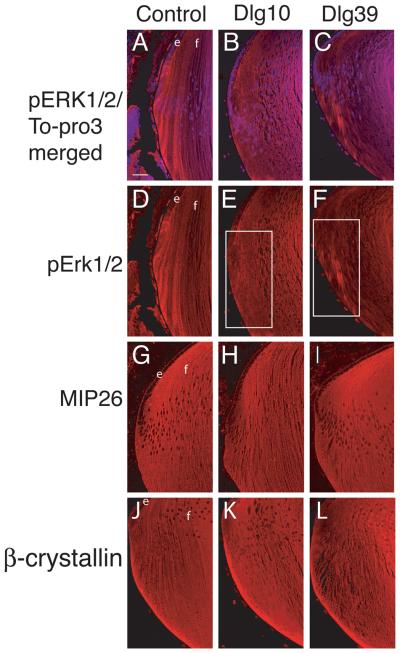
Figure 6. Reduced accumulation of pERK in transition zone of lenses from Dlg10 and Dlg39 mice.
Longitudinally oriented, paraffin embedded eye sections from P2 control (A,D,G,J), Dlg10 (B,E,H,K) and Dlg39 (C,F,I,L) mice were subjected to immunoflourescence analysis for three markers of fiber cell differentiation. A-C: Merged images of control (A), Dlg10 (B) and Dlg39 (C) lenses stained with To-PRO3 (blue nuclei) and an anti-pERK antibody (red). D-F: Unmerged images corresponding to A-C show anti-pERK staining (red) only. White boxes show the patchy and reduced accumulation of pERK staining in the transition zone in Dlg10 (E) and Dlg39 (F) mice. G-H: Sections from control (G), Dlg10 (H) and Dlg39 (I) mice were immunostained with anti-MIP26 antibodies. The pattern of MIP26 immunostaining was not altered in lenses of Dlg10 or Dlg39 mice as compared to controls. J-L: Sections of control (J), Dlg10 (K) and Dlg39 (L) mice were immunostained with anti-β crystallin antibodies. The pattern of β-crystallin immunostaining was not altered in lenses of Dlg10 or Dlg39 mice as compared to controls. e=epithelium, f=fibers. Scale bar=50μm.

