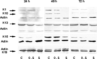
Fig. 2.
Analysis of expression of keratins by Western blot. Total protein was isolated from HaCaT cells after treatment with low (0.5 mM) or high dose (5 mM) of NaF during 24, 48, and 72 h and subjected to Western blotting. C, non-treated cells. Monoclonal antibodies LH1, 1C7, LHK 15, and b 170, respectively, against keratins 1/10, 13, 15, and 19 reacted with the expected bands at 56, 52, 62, 57, and 40 kDa, respectively. Anti-actin serum was added as control, eliciting a band at 40 kDa. Keratins 1/10 (K1/10) and 15 (K15) proteins were not detected after high dose of NaF treatment during the three times of observation. Keratin 13 (K13) and keratin 19 (K19) detection was not modified at any exposure time neither at low dose and high dose of NaF treatment

