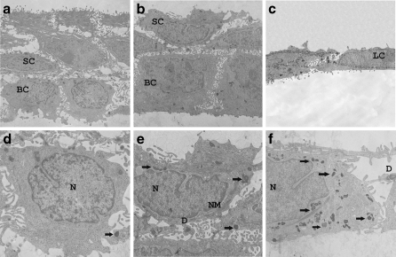
Fig. 6.
Stratification of HaCaT cells. HaCaT cells were treated at low (0.5 mM) and high (5 mM) of NaF for 3 weeks on a nitrocellulose membrane to achieve stratification. C, non-treated cells. In overviews, control (a) and low dose (b) membrane showed partial stratification with cubic basal cells and polygonal suprabasal cells. At high dose (c), HaCaT cells appear as monolayer. Detailed views (d–f) showed normal cells with intact nucleus (N), nuclear membrane (NM), and desmosomes (D). The cytoplasm of treated cells is normal with normal organelles (Golgi, ER, etc.; not shown), although there are granules (f, black arrows)

