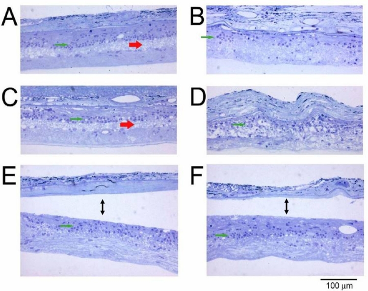
FIGURE 21.
Retinal sections from the right eye of a patient who died from an unrelated event, which was implanted with an ASR device. Sections shown in A through D are 1 to 2 mm from implant. Sections in E and F contain the implant area (between arrows). The red arrows indicate remnants of the inner plexiform layer. The green arrows point to remaining inner nuclear layer cells (toluidine blue–stained resin sections 160X).

