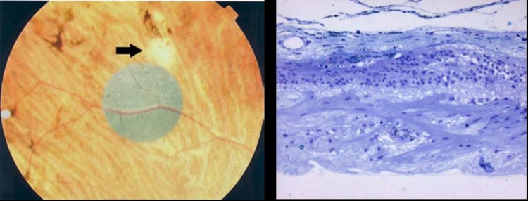
FIGURE 22.
Fundus photograph of the right eye of patient 3 showing the location of the ASR device. Note that white spot (black arrow) located superior to the ASR device appears to correspond to the histologic section shown on the right. The ganglion cell layer is composed of thickened fibrous tissue (toluidine blue–stained resin sections, 160X).

