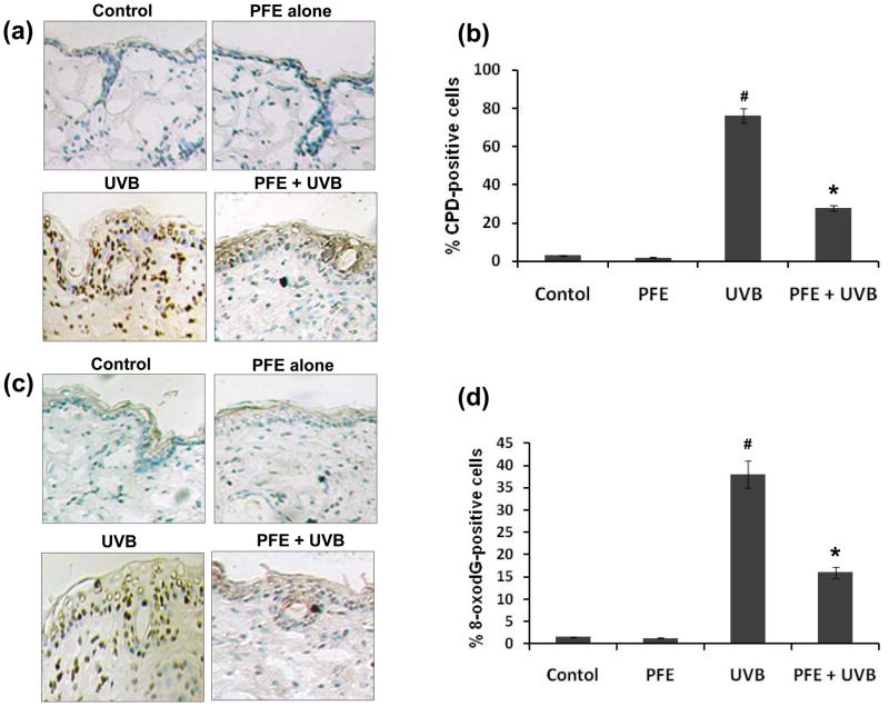
Figure 5. Inhibitory effect of PFE on UVB-induced formation of cyclobutane primidine dimers and 8-oxo-7,8-dihydro-2′-deoxyguanosine in SKH-1 hairless mice.
Twenty four hours post-UVB irradiation, the animals were sacrificed, skin biopsies were taken and frozen in OCT. Skin sections, 6 μm thick, were cut and processed for immunostaining. Immunohistochemical staining for CPDs [a], and 8-oxodG [c] was performed using appropriate antibodies. Representative pictures are shown. The number of CPD positive cells (b) and 8-oxodG positive cells (d) after immunostaining were counted in five different areas of the sections under a microscope. The numbers of CPD and 8-oxodG positive cells are represented as percent of CPD and 8-oxodG positive cells respectively. The data represents the mean ± SE of 8 mice (#p < 0.001 vs control; *p <0.001 vs. UVB).

