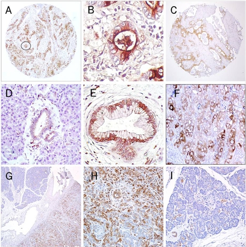
Figure 1.
Expression of CD74 in pancreatic tissues. (A) and (C) (50×) – an intense diffuse reaction for CD74 expression is noted within specimens of invasive pancreatic carcinoma with labeling of the apical cytoplasm and cell surface evident in (B) (circled field from A; 400×). (D) normal pancreatic acinar cells are mostly negative with weak, limited labeling of normal ductal tissue (200×). (E) PanIN-3 lesion shows evidence of a mostly cytoplasmic/perinuclear staining (200×). (F) Focal, CD74 expression within the CaPan1 human pancreatic carcinoma cell line grown as a xenograft in athymic nude mice (200×). (G) A specimen demonstrating chronic pancreatitis with fields of both involved and noninvolved tissue (50×); (H) the involved fields demonstrate CD74 staining of acinar and ductal cells (100×), whereas in I – only focal staining of ductal tissue is evident within the noninvolved field (100×).

