Figure 1.
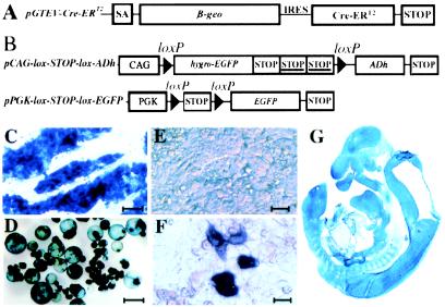
Generation of GTEV ES cells stably expressing pGTEV-Cre-ERT2. (A) Structure of the bicistronic gene-trap expression vector pGTEV-Cre-ERT2. SA, adenovirus major late-transcription splice acceptor; β-geo, LacZ–neomycin phosphotransferase fusion gene conferring resistance to G418 and expression of β-galactosidase; Cre-ERT2, gene encoding the ligand-dependent Cre recombinase Cre-ERT2; IRES, internal ribosomal entry site of the encephalomyocarditis virus; STOP, transcription termination from simian virus 40. (B) Structure of the pPGK-lox-STOP-lox-EGFP and pCAG-lox-STOP-lox-ADh reporter plasmids. CAG, cytomegalovirus/chicken β-actin fusion promoter; PGK, promoter of the mouse pgk-1 gene; STOP, transcription termination from simian virus 40; STOP, transcription termination from the mouse pgk-1 gene. (C and D) Histochemical analysis of β-galactosidase expression in undifferentiated ES cells from clone GTEV49 (C) and in 14-day-old embryoid bodies made from GTEV49 ES cells (D). (E and F) Histochemical analysis of Cre activity in undifferentiated ES cells from clone GTEV49. To detect Cre activity, GTEV49 ES cells were transfected with the pCAG-lox-STOP-lox-ADh reporter plasmid in the presence of 1 μM 4′OHT (F) or in vehicle alone (E). Cells were harvested after 48 h and processed for detection of ADh activity. (G) Histochemical analysis of β-galactosidase expression on a sagittal section of transgenic fetus tgGTEV49 at 13 days of gestation. [Scale bars = 10 μM (E and F) and 100 μM (C and D).]

