Figure 1.
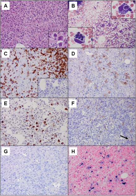
A. Hematoxylin and eosin (H&E) of bone marrow biopsy with marrow space virtually entirely replaced by the neopiastic process and hemophagocytosis (400×); insert: neopiastic large cells (1000×). B. Wright stained aspirate smear with trilineage hematopoiesis and no neopiastic large cells (100×); insert: hemophagocytic macrophages (1000×). C. CD3 stain demonstrating surface positivity on the neopiastic large cells as well as numerous background small T-cells (400×); insert: PAX-5 stain negative on the large cells (400×). D. CD30 stain demonstrating membrane and Golgi uniform positivity on the neopiastic large cells (400×). E. Granzyme B stain demonstrating positivity on the neopiastic large cells (400×). F. CD56 stain demonstrating positivity on scattered small background lymphocytes as well as faint staining on rare lymphoma cells (arrow) (400×). G. ALK-1 stain negative on lymphoma cells (400×). H. EBER in situ hybridization demonstrating strong uniform positivity on the neopiastic large cells (400×).

